Scanning transmission X-ray microscope (STXM) principle is a simple one. [1] A zone plate focuses X-ray beam into a small spot, a sample is scanned in the focal plane of the zone plate and the transmitted X-ray intensity is recorded as a function of the sample position. STXM name is usually used for microscopes working in soft X-rays range. In order to achieve time-resolved imaging of the magnetisation dynamics of a certain magnetic configuration at a STXM, we employ a stroboscopic scheme where the excitation is the pump and the synchrotron x-ray flashes are the probe.
Absorption of circular or elliptically polarized X-rays depends on interaction of this beam and magnetic moment of the absorbing atoms [2]. The effect is called X-ray magnetic circular dichroism (XMCD). The sample can be tilted by 30° with respect to the X-ray beam, allowing for measuring the in-plane component of the magnetization.
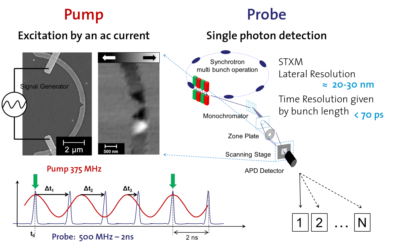
Figure 1: Illustration of time-resolved scanning transmission x-ray microscopy. The imaging is performed in a stroboscopic way by delaying the electronic excitation of a magnetic structure (pump) relative to the x-ray flashes (probe). Single Photon detection allows for multi-channel acquisition, which can be performed in standard multi-bunch operation mode of the synchrotron.
Beamlines:
Molecular Science Beamline 11.0.2 [3], Advanced Light Source (ALS), Berkeley CA, USA
Magnetic X-Ray Micro- and UHV-Spectroscope (MAXYMUS), BESSY, Berlin, Germany
References:
[1] Technical report "STXM in nanoscience", Tolek Tyliszczak and Kai Wei Chou.
[2] G. Schütz et al., Phys. Rev. Lett. 58, 737 (1987).
[3] A. L. D. Kilcoyne et al., J. Synchrotron Radiat. 10, 125 (2003).